Diagnosis and Treatment of Macular Degeneration
Diagnosis of Macular Degeneration
Like many eye conditions and diseases, it is often possible to detect early signs of Macular Degeneration during your regular eye examination. Scheduling regular eye exams is an excellent way for Dr. Whitaker to make an early diagnosis of Macular Degeneration.
Equally as important as a regular eye examination is being aware of the symptoms that may indicate the early presence of Macular Degeneration. If at anytime you experience any “distortion” or “twisting”, “shadowing” or “bending” of objects in your vision, you should schedule an appointment at Riverside Eye Center immediately. Be sure to let the receptionist know that you are experiencing these symptoms.
If you are over the age of 40-45 and you have a family history of Macular Degeneration, Dr. Whitaker recommends that you have a thorough eye examination, including a dilated retinal evaluation, each year. Please be sure to tell our staff if you have a family history of Macular Degeneration.
During your eye examination, eye drops will be put in your eyes to dilate your pupils in order to carefully examine the Macula and Retina using various types of instruments and sources of high magnification.
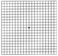
Additional tests that Dr. Whitaker may perform to further evaluate the Macula during your eye examination can include checking your color vision and an Amsler Grid Test. The Amsler Grid Test helps identify distortion of your central vision, and may be a subtle indication of swelling or fluid under the Macula.
The Amsler Grid Test  Although the Amsler Grid Test is relatively simple, it is very useful in detecting small changes in your vision that can result from the accumulation of just a minimal amount of fluid under your Macula. Dr. Whitaker may ask you to take an Amsler Grid home and use it each day to check for slight changes in your vision. If this is necessary, Dr. Whitaker and staff will supply you with an Amsler Grid and detailed instructions on how to use it. If, during your examination, Dr. Whitaker detects any signs of Macular Degeneration or he believes that you may be at risk for Macular Degeneration, he may schedule for additional testing.
Fluorescein Angiography
In order to effectively diagnose Macular Degeneration, Dr. Whitaker may find it necessary to take detailed color photographs of your Macula and Retina. It may also be necessary for you to have a Fluorescein Angiogram (FA), also called an Intravenous Fluorescein Angiogram (IVF). The IVF is performed right in the comfort and convenience of our office at Riverside Eye Center in order to study the health and circulation of the Macula and Retina.
To prepare you for an IVF, eye drops will be placed in your eyes to dilate your pupils. Then a fluorescent dye, called Sodium Fluorescein, is injected into a vein in your arm. The dye will begin to circulate after about 10-15 seconds at which a series of photographs will be taken in rapid succession using a high speed digital retinal camera as the dye passes throughout the retinal blood vessels. From these pictures, if present, Dr. Whitaker will be able to see any fluid leakage or new blood vessel growth beneath the Retina. The IVF will also show any changes or damage to the Macula and Retina and the extent of the changes. Most important, Intravenous Fluorescein Angiography gives Dr. Whitaker a great deal of information regarding whether certain types of treatments such as Avastin Injections, Eylea Injections or Lucentis Injections might help stabilize your vision and prevent vision loss. Today, thanks to the advances in treating Wet Macular Degeneration, if caught early, it may be possible to avoid suffering significant vision loss.
Treatment of Macular Degeneration
If Macular Degeneration is diagnosed early enough, we are very fortunate to have a number of possible treatment options that may help to slow or even halt the progression of vision loss from Macular Degeneration. However, patients must understand that once the Macula has been damaged, there is no treatment that currently can reverse that damage and the associated loss of vision. Early diagnosis and treatment to prevent or halt vision loss must be the approach that we take.
Macular Laser Photocoagulation
The National Eye Institute has sponsored a large scale multisite clinical trial in order to determine what particular macular conditions should be treated with lasers, what types of lasers should be used, which patients might get the best results and to try and establish the best ways to use lasers to treat macular degeneration. Dr. Whitaker routinely reviews results from studies such as The Macular Photocoagulation Study (http://www.nei.nih.gov/neitrials/viewStudyWeb.aspx?id=60) in the hope of finding a set of useful clinical guidelines for the Laser Treatment of Macular Degeneration. Unfortunately, the overall findings of the Macular Photocoagulation Study suggest that it is limited in its effectiveness and may also lead to scarring of the Macula and additional vision loss. As such, except in rare cases, Dr. Whitaker does not perform Macular Laser Photocoagulation Treatment for Macular Degeneration.
Visudyne Photodynamic Laser Therapy
Another type of Laser Treatment for Wet Macular Degeneration uses a light-activated drug called VisudyneTM. Visudyne works through a “cool” process that produces a selective destruction of the weak leaky new blood vessels that grow under the Macula. The purpose of the Visudyne Photodynamic Laser Treatment is to seal off leaking vessels while leaving healthy ones intact. Unfortunately even when successful, Visudyne Photodynamic Laser therapy does not always prevent recurrence of the new blood vessel growth. It is often necessary to have repeated treatments in order to slow the progression of vision loss, and even with repeated treatments a recurrence of neovascularization is possible and must be carefully monitored to preserve vision. If you are a candidate for Visudyne, Dr. Whitaker will fully discuss its risks and benefits.
Vascular Endothelial Growth Factor Inhibitors (VEGF)
Avastin, Eylea and Lucentis Injections
As a result of advanced cancer research in the area of “angiogenesis” or new blood vessel growth, considerable information has been obtained in the treatment of Wet Macular Degeneration. Researchers discovered that a specific protein called “Vascular Endothelial Growth Factor” (VEGF) causes the growth of new blood vessels or “neovascularization” to occur in the eye. From this work, drugs that can be injected into the eye in order to slow or stop the growth of new blood vessels. Two drugs, Eylea and Lucentis have been developed and FDA approved with specific indications to treat Wet Macular Degeneration and one drug, Avastin has been FDA approved with an indication for the treatment of metastatic colorectal cancer. However, Once a drug is approved by the FDA, physicians may use it “off-label” for other purposes if they are well-informed about the product, base its use on firm scientific method and sound medical evidence, and maintain records of its use and effects. Many Ophthalmologists, including Dr. Whitaker, now use AvastinTM “off-label” to treat Age Related Macular Degeneration and other eye conditions that cause neovascularization, since research indicates that VEGF is one of the causes for the growth of the abnormal vessels that cause these conditions. Each of these drugs works by inhibiting Vascular Endothelial Growth Factor (VEGF) so that there is little or no stimulus to grow new blood vessels in the Retina.
Avastin, Eylea and Lucentis Injections are intravitreal injections-that means an injection that is placed directly into the Vitreous of the eye. Generally they need to be repeated every four to six weeks. Clinical studies of these anti-VEGF Injections indicate that when given to patients who have evidence of new blood vessel formation monthly, over 90% of patients will maintain their vision (http://www.fda.gov/).
Should you be at risk for Wet Macular Degeneration, Dr. Whitaker will discuss more about the results with you. He will also be able to tell you more about the length of your actual treatment program, as it varies for each patient. If Avastin Injection, Eylea Injection or Lucentis Injection is a possible option for you, Dr. Whitaker will spend the time necessary to thoroughly review the possible risks, benefits and side effects with you before you decide to proceed.
Age Related Macular Degeneration & Diet
It is believed that nutrition may play a role in the likelihood of developing Macular Degeneration. Studies indicated that people who have a diet that is rich in fruits and vegetables-particularly green leafy vegetables-have a considerably lower incidence of Macular Degeneration. It is not certain whether taking dietary supplements can prevent progression in patients with existing macular disease, but it does seem clear that certain dietary supplements can reduce your risk of Macular Degeneration. The Age Related Eye Disease Study (AREDS), which was sponsored by the National Eye Institute (http://www.nei.nih.gov/amd/summary.asp), showed that taking high levels of antioxidants and Zinc could reduce the risk of developing Age Related Macular Degeneration by about 25%. This is not a cure, but we need to consider this information as a possible way to help patients who are at risk for Age Related macular Degeneration prevent vision loss.
NOTE:A VERY SPECIFIC FORMULATION WAS USED IN THIS STUDY
BEFORE patients begin taking any course of vitamin or antioxidant supplements, you should fully discuss the risks and benefits with Dr. Whitaker, who in consultation with your family physician or Internist, will determine whether this is safe and effective for you to try.
